A clearer view inside of people
A cyclist has a serious accident, and is transported as quickly as possible to a hospital. Now the doctors must swiftly decide: How complicated is the injury? Is there spinal damage? Are important blood vessels affected?
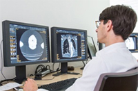
Answering these questions requires the experienced eye of a radiologist who can quickly evaluate CT images, so that doctors from the various departments can immediately begin treatment.
Within a matter of seconds, CT scans generate precise images of organs, bones and the brain, and just minutes after an examination our radiologists are provided with up to 5000 images. Radiologists then need to consult with surgeons to diagnose what needs immediate attention and which treatments are required. At the Medical Center – University of Freiburg, radiologists deal with emergency patients in the University Emergency Center (UNC), and also examine regular patients every day in the Department of Radiology.
Minimal radiation exposure
Besides CT scans, other important imaging techniques include magnetic resonance imaging (MRI), X-rays, mammography and angiography. “An MRI is the first choice for oncological patients. Using magnetic fields and radio waves allows us to effectively diagnose and classify tumors,” says Professor Dr. Mathias Langer, Medical Director of the Department of Radiology. Both CT scans and X-rays radiate the body. X-rays allow radiologists to image bone fractures in two dimensions while CT scans are better equipped to diagnose more complicated bone fractures. “At the Medical Center – University of Freiburg, we use state of the art equipment and pay special attention to radiation dosage in order to keep patient exposure to radiation as low as possible,” emphasizes Professor Mathias Langer.
Mammography, an X-ray imaging procedure for examining the breast, also minimizes patient radiation exposure and is always complemented by an ultrasound test. A further type of breast examination called tomosynthesis uses compressed layers of images to more precisely picture masses in the female breast. An MRI is another imaging technique which can be used for breast examinations as necessary.
Angiography is a technique which uses X-rays to image the inside of blood vessels and detect constricted vessels or internal bleeding. To perform this procedure, radiologists insert a catheter into the patient’s thigh, through which a guide wire or a tube is fed to reach the point of care. A contrast agent is injected into the blood vessel there, allowing an X-ray image of the inner vessel to be generated.
Back