More precise, faster and more comfortable: Germany's most state-of-the-art PET/CT device comes into service
New device offers optimized diagnostics, improved patient comfort and reduced radiation exposure / Combination of positron emission tomography (PET) and computed tomography (CT) is important, among other things, in cancer diagnostics.
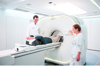
In patients with cancer, a PET/CT scan is often performed to detect the exact location and metastases of a tumor. The latest device of its kind, in the Department of Nuclear Medicine at the Medical Center - University of Freiburg, enables doctors via its all-digital technology to perform examinations with high spatially detailed accuracy and the least possible stress for patients. With the help of these images, diseased tissue can be precisely localized and the disease stage better determined. Based on this, individually tailored treatment plans can be defined and their effectiveness monitored as well. The compact design of the "Vereos Digital PET/CT" scanner from Philips, with a wider device opening, reduces the sense of tightness, so that even patients with claustrophobia can be examined. Moreover, special lighting effects and projections on the wall of the examination room contribute to a relaxed examination atmosphere.
"With the new device we achieve a higher image resolution and at the same time can more than halve the duration of an examination as well as the radiation exposure," says Prof. Dr. Dr. Philipp T. Meyer, Medical Director of the Department of Nuclear Medicine at the Medical Center - University of Freiburg. In PET, the body is exposed to approximately the same amount of radiation as an annual natural radiation dose. "In view of the high diagnostic benefit, this can be regarded as quite safe, which is for example very important in follow-ups and in examining children and adolescents," says Prof. Meyer. A PET visualizes those metabolic processes and signaling pathways in the body that are already altered in the early stages of tumors and other diseases. This "functional" or "molecular" imaging is complemented by CT, in which X-rays depict organ and bone structures in high resolution. Using the superimposed images from both imaging techniques, called image fusion, creates a three-dimensional map of the body on which diseased tissue emerges as glowing dots. Doctors can thus plan individualized operations as well as chemo- or radiotherapy. "The goal is to find a personalized, optimal treatment for the patient with the least side effects," says Prof. Meyer.
A further major advantage of the examination is the whole-body diagnostics with both PET and CT. "Because we perform two procedures at the same time, we spare patients the time and travel involved in duplicate examinations. The practitioners also get the results faster. In addition, many studies show that the combined examination is more accurate and helpful than the sum of individual ones," says Prof. Meyer. The scanner's high-resolution whole-body images take only about 15 minutes. The scanner is about 500 kilograms lighter than conventional systems. Without the table, the device measures just 1.30 meters and has been condensed into a much shorter and more compact system. Instead of two ring openings for CT and PET, in this latest scanner there is only one ring. In addition to oncological issues, the PET/CT examination is also used successfully in other, for example, rheumatological, cardiological or neurological diseases.
PET examination background: PET is a three-dimensional imaging technique that maps biochemical functions such as an increase in metabolism or changes in signaling pathways in the body. By means of a radioactive substance which is injected into the patient - so-called tracers or radiopharmaceuticals - numerous tumors and their metastases in the body can be made visible.
CT examination background: The CT is used in X-ray diagnostics and works with X-rays. The cross-sectional images from inside the body provide information about the location, size and spread of, for example, diseased tissue.
*Please note that this examination can only be performed if recommended by a doctor.
Back