Fast, precise and gentle: innovative cardiac imaging in Freiburg
A new device at the Medical Center - University of Freiburg delivers high-resolution images of the heart. At the same time, patients' safety and comfort are significantly increased. It is used mainly in the diagnosis of chronic and acute heart problems.
Recently, the Department of Nuclear Medicine at the Medical Center - University of Freiburg has been using a highly sensitive cardiac SPECT device that is available at only a few locations in Germany. It is the first device of its kind in Baden-Württemberg. It allows the circulation and contraction of the heart to be determined much faster and more precisely. A session lasts at most ten minutes, less than half as long as comparable methods. The system is intended for the diagnosis and monitoring of chronic heart problems, but due to the swiftly available findings it can also be employed for new or acute symptoms.
"The new device improves on non-invasive diagnostics for patients with suspected or already established circulatory disorders of the heart. The examination is significantly faster and more comfortable and the image quality is much better. At the same time we can reduce the required radiation dose below the annual natural radiation exposure level. As a result, examination with the new device is also gentler compared to other methods such as cardiac catheterization and computed tomography," says Prof. Dr. Dr. Philipp T. Meyer, Medical Director of the Department of Nuclear Medicine at the Medical Center - University of Freiburg.
What is a cardiac SPECT exam and how does it work?
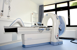
SPECT stands for "single-photon-emission computed tomography". In a SPECT scan, a slightly radioactive substance is first injected into a brachial vein, and spreads quickly with the blood through the body. Depending on the substance used, particular body and organ functions can be made visible. With the SPECT camera, the emitted radiation is detected and used to create sectional images. For example it is possible to examine bones in tumor diseases or orthopedic problems, brain functions in neurological diseases such as Parkinson's or, in the case at hand, circulatory disorders of the heart. The "Discovery NM 530c" device used at the Medical Center - University of Freiburg has been optimized especially for cardiac SPECT examinations. The device has nineteen independent cameras that simultaneously take pictures of the heart from different angles. Thanks to special semiconductor technology, this is much more sensitive and faster than conventional systems.
Also suitable for patients with claustrophobia
In addition, examinations with the new, more compact system are considerably more convenient, and are no problem for patients with claustrophobia. Circumstances under which other methods cannot be used, such as contrast agent allergy, kidney or thyroid disease, or pacemakers, also present no obstacles to this procedure.
When is a cardiac SPECT scan performed?
In a cardiac SPECT exam, circulatory disorders of the cardiac muscle can be detected both at rest and under physical or medicinal stress. It is used in addition or as an alternative to invasive methods such as cardiac catheterization or other imaging techniques. During the examination, the contraction processes of the heart are also recorded, which provides important information about the heart's pumping power. If needed, the nerves supporting the cardiac muscle can also be examined.
"We are delighted to now have these diagnostics quickly and easily available in Freiburg. It represents a diagnostic gain and a great relief for many of our patients", says Prof. Dr. Dr. h.c. Christoph Bode, Medical Director of the Department of Cardiology and Angiology I at the Heart Center, University of Freiburg - Bad Krozingen.
Back