Prostate Cancer Diagnostics Using PSMA-PET/CT

©Medical Art Inc / istock
[27.12.2022] In recent years, PET/CT with ligands of the prostate-specific membrane antigen (PSMA) has become an established imaging technique for state-of-the-art diagnosis of prostate cancer. The Department of Nuclear Medicine at the Medical Center - University of Freiburg has been involved in the development of PSMA-PET/CT for more than 10 years.
In the situation of tumor recurrence (e.g. after surgery or radiation therapy), PSMA-PET/CT is well-established and essential for detection and localization of tumor recurrence and possible metastases as well as subsequent planning of further treatment (e.g. salvage lymphadenectomy or radiotherapy) according to current guidelines.
However, PSMA-PET/CT is also highly valued in newly diagnosed prostate cancer: The recently published proPSMA Study showed a significant superiority of PSMA-PET/CT compared to the conventional diagnostic algorithm with contrast-enhanced CT and bone scintigraphy in patients with a so-called high-risk constellation. Preoperative PET/CT led to a change of the treatment plan in 28% of cases (i.e. twice as often as standard diagnostics, 15%) in order to achieve optimal treatment results and cure of patients. In the case of detection of unexpected metastases, for example, a systemic (drug) treatment instead of originally planned surgery or radiation therapy was chosen in 15% of patients. Thus, optimal staging examination using PET/CT enables a personalized treatment that is most appropriate for an individual patient to reduce the risk of tumor recurrence and treatment side effects.
The Department of Nuclear Medicine at the Medical Center - University of Freiburg has been involved in the development and validation of PSMA-PET/CT for more than 10 years. To ensure optimal examination quality, fluorine-18-labelled PET tracers and modern, fully-digital PET/CT scanners are used.
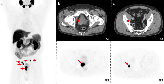
PSMA-PET/CT ©UKF PSMA-PET/CT with [18F]PSMA-1007 in a 71-years-old prostate cancer patient with high-risk constellation (Gleason score 8). The three-dimensional view of the body (a) and horizontal PET/CT cross-sections show intense PSMA expression of the primary tumor in the prostate (b) and local lymph node metastases (c) (see arrows). Based on this information, curatively intended surgery can be optimally planned.
PSMA radioligand therapy
A very promising and highly effective therapeutic complement to PSMA-PET/CT diagnostics is the PSMA radioligand therapy, which is very well tolerated due to its targeted mode of action. Patients with advanced tumor stages receiving this therapy are treated with great success at the Department of Nuclear Medicine at the Medical Center - University of Freiburg.
Prof. Dr. Dr. Philipp Tobias Meyer, medical director of the department of Nuclear Medicine at the Medical Center – University of Freiburg
Prof. Dr. Juri Ruf, executive senior physician of the department of Nuclear Medicine at the Medical Center – University of Freiburg
____________________________________
Literature:
- Hofman MS, Lawrentschuk N, Francis RJ, et al. Prostate-specific membrane antigen PET-CT in patients with high-risk prostate cancer before curative-intent surgery or radiotherapy (proPSMA): a prospective, randomised, multicentre study. Lancet. 2020;11;395(10231):1208-1216.
- Oncology guideline program (German Cancer Society, German Cancer Aid, AWMF): S3 guideline prostate cancer, long version 6.2, 2021, AWMF registry number: 043/022OL, www.leitlinienprogramm-onkologie.de/leitlinien/prostatakarzinom/ (retrieved on: 11/03/2022).