Identifying breast cancer more reliably
[08.01.2017] New technology allows more exact breast tissue images without additional radiation exposure / False-positive findings are minimized.
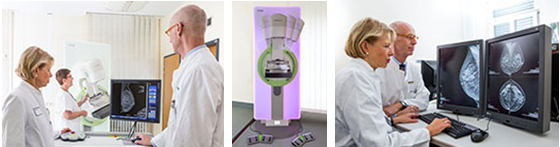
© Medical Center - University of Freiburg/Britt Schilling
Breast cancer is the most common malignant tumor in women. Every year, 70,000 new cases are registered in Germany alone. As a rule, the chances of recovery increase the sooner the tumor is discovered and the more accurately it can be diagnosed. In the gynecological radiology department of the Department of Radiology at the Medical Center - University of Freiburg, a new X-ray technique has recently become available for the first time in Central Europe. It represents a considerable development in diagnostic safety, without significant additional radiation exposure.
For an optimal diagnosis, conventional 2D mammography images of the breast tissue are combined with 3D tomosynthesis displays. "Thanks to the 3D display, one can determine with great certainty whether condensations in the 2D image are due to tissue overlay or in fact to a carcinoma. In this way, false-positive findings can be minimized, "says Prof. Dr. Mathias Langer, Medical Director of the Department of Radiology.
In the Siemens Mammomat's new high-definition tomosynthesis, the X-ray tube swings around the breast in a 50-degree arc, making 25 individual images of the breast tissue, with very low X-ray exposure each time. From these two-dimensional images, high-resolution 3D images are subsequently reconstructed. "While generating the findings, we can move on the monitor through the individual layers of breast tissue. Thanks to newly developed calculation methods, the image quality is significantly increased, so we can recognize microcalcifications more clearly, and more easily identify changes as benign or malignant," explains Prof. Langer.
In addition to the improved diagnostic possibilities, the new technology also helps to reduce radiation exposure. For reliable assessment, especially in case of a dense mammary gland body, two 2D mammographs from different planes are required in addition to 3D tomosynthesis. If previously a total of three X-ray images had to be taken, now one of the two 2D mammographies needed for high-definition tomosynthesis can be used. So one of the conventional 2D mammographies can be dispensed with, and the entire X-ray dose reduced by about 20 to 30 percent. "This allows us to use all the technologies without the patient having significantly increased radiation exposure," says Dr. Marisa Windfuhr-Blum, the responsible senior physician in gynecological radiology at the Medical Center - University of Freiburg. In combination with a clinical as well as an ultrasound examination, the new equipment thus allows a significant improvement in diagnostics.
See also: