Lungs in Good Hands
(Part I)
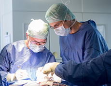
The lung is a vital organ. In the Department of Thoracic Surgery, malignant diseases such as lung cancer and lung metastases are carefully diagnosed and treated using minimally invasive surgical procedures.
___________________________
Insights into the airways
If diseases of the respiratory tract and lungs are suspected, the central air passages (the trachea and large bronchi from the first to the second branches) can be examined using a bronchoscope. Usually a flexible optical system is used, which transfers images directly to a monitor via a video chip. Tissue samples can taken through a working channel using small forceps and brushes, and secretions can be removed using suction. The examination is not painful, the only unpleasant part being a more or less pronounced cough. Therefore, before and during the examination a local anesthetization of the mucous membrane is performed, and optionally an additional light sleeping agent is administered. In an endobronchial ultrasound examination (EBUS), an ultrasound head is integrated into the bronchoscope. In this way the lymph nodes in the thoracic cavity can be examined quite gently. These play an important role in lung cancer therapy. "Additional important pathological examinations such as mutation analyses are also now possible without any big difficulties," says Dr. Mirjam Elze, senior physician in the Department of Thoracic Surgery at the Medical Center - University of Freiburg. An additional mediastinoscopy for enlarged lymph nodes is only necessary in case of insufficient results from the puncture.
Electromagnetic navigation
The physicians in the Department of Thoracic Surgery use the electromagnetic navigation method In order to reach small pulmonary nodules and to gently but purposefully probe into the finest branches of the bronchi. In preparation for the bronchoscopy, the patient's CT data are used to plan for and simulate the examination. The target point is marked for this purpose. During the examination the patient lies inside an electromagnetic field. A specialized probe is inserted using the bronchoscope, the tip of which can be localized electromagnetically. During the bronchoscopy the system guides the examiner and bronchoscope, similarly to GPS navigation, to the previously defined target points. While doing this, the examiner constantly receives a three-dimensional localization image and an indication of the distance to the target, where tissue samples can then be removed for diagnosis. "In the future, it will also be possible to treat small malignant pulmonary nodules with steam or microwaves, thus sparing the patient from surgery," says Dr. Elze.
Bronchoscopy and thermal procedures
In the Department of Thoracic Surgery so-called thermal procedures are used when hemoptysis occurs, and above all in case of malignant processes in the area of the respiratory tract, leading to displacement of the trachea or bronchial tubes with resulting air dysfunction. These include argon plasma coagulation, laser- and cryotherapy. Devices are used for this which superficially scab, freeze or vaporize the tissue. In this way the air passages can be re-opened and the symptoms can be significantly alleviated.
Alleviate dyspnea with a stent
If a tumor threatens to block the air passages, a stent can allow freer breathing. This is a lattice framework in the form of a tube of metal or plastic which keeps the airways open. "This alleviates or even completely eliminates the patient's feeling of breathlessness and suffocation," says Dr. Elze. The most commonly used stents today are made of silicone or nitinol, a nickel-titanium alloy, and can be removed if needed.
See also: Department of Thoracic Surgery
Back