Uterine Fibroids Embolisation
Uterine fibroids are benign (non-cancerous) tumours which develop on or within the muscular wall of the uterus. They are comprised of dense, fibrous tissue which are fed by blood vessels. Fibroids are frequent occurrences and are the most common benign tumours in women. Doctors estimate that fibroids become clinically relevant in about 25 % to 30 % of all women aged between 30 and 50 years. Uterine fibroids can grow in various parts of the uterus.
Intramural fibroids are most common. Because these fibroids grow in the muscular wall of the uterus, they make it feel larger than normal and can cause an increase in menstrual bleeding, pelvic pain, back pain or pressure.
Subserosal fibroids are the second most common. Because these are located on the outer wall of the uterus, they do not usually affect menstrual flow. However, they can cause pelvic pain, back pain or pressure.
Submucosal fibroids can cause heavy or prolonged periods, even if they are very small.
Typically, women who have uterine fibroids have more than one fibroid and they can vary in size. Some are no bigger than a pea while others can grow to the size of a melon. When fibroids are diagnosed, the extent of the disease is determined by comparing the size of the uterus to a typical size during pregnancy. For example, a large fibroid or multiple fibroids may enlarge the uterus to the same size as a six or seven month pregnancy.
The exact reason why uterine fibroids develop is unknown. However, medical researchers have associated the condition with several factors – e.g. age, genetics and hormones. There is a strong genetic component to fibroid development, which causes fibroids to occur at least three times more frequently among black women.Uterine fibroids can dramatically increase in size during pregnancy. It is thought that this effect is due to the increase in the amount of oestrogen – the female hormone – that naturally occurs during pregnancy. After delivery, the fibroids usually shrink to the size they were before the pregnancy. During menopause oestrogen levels dramatically decrease. This causes uterine fibroids to shrink, relieving symptoms. However, if a women takes hormone replacement therapy (HRT) during menopause, oestrogen levels do not decrease, the fibroids may not shrink and the symptoms may remain.
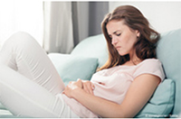
TYPICAL SYMPTOMS
- Heavy, prolonged menstrual periods and unusual monthly bleeding –
- sometimes with clots – which can cause anaemia
- Increased menstrual cramping
- Pain, pressure or discomfort in the pelvis
- Pain in the back, sides or legs
- Pain during sexual intercourse
- Blockage of urine flow from the kidney to the bladder
- Urinary frequency due to pressure on the bladder
- Constipation and/or bloating due to pressure on the bowel
- Abnormally enlarged abdomen
Your doctor may diagnose fibroids during a routine gynecologic examination. In order to confirm this diagnosis, your doctor may request that you undergo an ultrasound or MRI (Magnetic Resonance Imaging) examination.
The treatment for uterine fibroids depends on the size and location of the fibroids and the severity of your symptoms. If you do not have symptoms, your doctor may decide that there is no need to treat the fibroids. However, your physician will probably recommend yearly visits to have them checked. If you do develop symptoms there are a number of treatment options available including:
Medical Therapy
Medical therapy for uterine fibroids may include the use of drugs to control symptoms.These drugs include nonsteroidal anti-inflammatory drugs (NSAIDs), birth control pills and hormone therapy (GnRHa).
Surgical Therapy
There are two surgical options for uterine fibroids – myomectomy and hysterectomy.
A Myomectomy removes uterine fibroids, without removing the uterus. Depending on the location of the fibroids, the myomectomy can be done through the pelvic area or through the vagina and cervix.
A Hysterectomy is a surgery performed to remove a woman’s uterus and cervix. A supracervical hysterectomy only removes the uterus. In all cases, menstruation stops and a woman loses the ability to bear children.
Non-Surgical Therapy – Embolisation
Uterine fibroid embolisation (UFE) is a less invasive approach that is designed to preserve the uterus. The embolisation procedure is carried out by an interventional radiologist who is especially trained to use image guidance methods to gain access to vessels and organs and treat many conditions. In general the embolisation procedure is performed while the patient is conscious but sedated, drowsy and feeling no pain. A small opening measuring 1.5 mm in diameter is made in the groin, through which a thin catheter is inserted into the artery. The catheter is guided through the arterial tree to the uterus while the radiologist watches the progress of the procedure using a moving x-ray (fluoroscope).Tiny microspheres, the size of grains of sand, are injected into the artery that is supplying blood to the fibroids, cutting of the blood flow. It may be necessary to repeat embolisation for fibroids on the opposite side, through the same opening and using the same catheter and microcatheter combination.
The average recovery time before patients return to work or their normal daily activities is approximately one week. The reason most patients are not able to return to work sooner is due to the post-embolisation syndrome which causes a light flu-like illness. Your gynecologist and interventional radiologist will take care of follow-up including a MRI or ultrasound exam in three to six months to assess the results of embolisation and to insure that the blood supply to the fibroids is eliminated. Over time, the fibroids shrink, relieving symptoms.
Back