Rare surgery at Medical Center - University of Freiburg
A 61-year-old patient with a pronounced lower jaw growth, presented himself in the Department of Oral and Maxillofacial Surgery at the Medical Center - University of Freiburg. About 55 years earlier, a partial resection in his upper jaw region had taken place. The feature had grown slowly since then, with further surgery rejected for fear of complications. The renewed presentation came about due to increasing difficulty in turning his head.
Wolfgang P. had his first facial surgery at the age of five. His jawbone had grown uncontrollably. Later, during adulthood, the bone simply continued to grow, until it was so large that it simply couldn't work anymore. Only then did Wolfgang P. undergo surgery at the Medical Center - University of Freiburg. The operation was successful in relieving him of a rare, benign, yet massive, four-kilo tumor. And also of its – in the truest sense of the word – burdensome side effects.
This patient had extensive fibrous dysplasia that had developed over many years. Due to the size of the growth, a complex virtual 3-D plan to prepare resection templates for the anatomically correct modelling of the lower jaw was necessary. Fibrous dysplasia is a congenital ossification disorder. When the facial skull bones are affected, clinically increasing asymmetries become evident. As treatment, modelled osteotomy is recommended, especially with extensive findings.
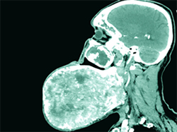
Clinically, there was an approximately 30 cm x 28 cm x 15 cm bone-hard mass enclosing three-quarters of the lower jaw. It was planned to ablate the growth via mandibular reshaping using a 3-D statistical model, which helped to determine the original shape of the lower jaw. In the CAD/CAM (computer-aided design/manufacturing) procedure, four resection templates were produced to implement the planned osteotomy intraoperatively. In case of extensive findings, computer-assisted surgery (CAS) based on precise preoperative 3-D planning can help to perform the modelled osteotomy predictably. As a virtual planning aid for unilateral (one-sided) findings, the established procedure is mirroring, in which the healthy side is mirrored onto the affected side. For bilateral findings, a more complex method must be used, in which a statistical model helps to calculate the patient-specific original form.In the present case, this technique was used to compute the shape of the original undeformed lower jaw. On the basis of this surface assessment, a total of four patient-specific resection templates were created in order to enable precise, swift implementation of the preoperative plan intraoperatively, while protecting anatomically sensitive structures.
The histopathological findings report revealed tissue weighing over three kilograms. In the postoperatively prepared digital volume tomography (DVT), the newly modelled lower jaw was shown to have its anatomically correct shape.
The postoperative course was normal. After seven days the patient was discharged into outpatient care, to present himself every six months for follow-up treatment.
Fibrous dysplasia is a genetically-based, sporadic bone disease, first described in 1942 by Lichtenstein and Jaffé, resulting in a disordered proliferation of osteoblasts that subsequently cannot form complexly-structured lamellar bones. Usually only one bone is involved, with the upper jaw being affected somewhat more frequently than the lower jaw. The polyostotic (multiple bone) form is altogether rarer. Growth usually stops after the end of puberty.
Anamnestically, an increasing, painless swelling of the face in often young patients is described. In very rare cases, malignant degeneration may occur, so long-term follow-up monitoring is recommended. Until the end of puberty, treatment should be primarily observational. With increasing asymmetry or functional impairment, a modelled osteotomy can be performed. In rare cases the operation can lead to a relapse with increased growth tendency.
Back