Positron Emission Tomography (PET)

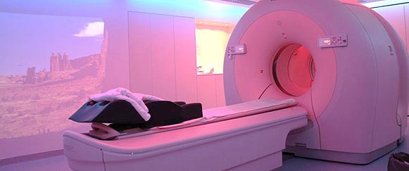
Modern PET/CT-system at our department (Philips GEMINI TF PET/CT)
PET is a unique tomographic examination that, unlike computer tomography or magnetic resonance imaging (CT or MRI), provides detailed information about body functions.
PET provides high-resolution imaging that was unknown to nuclear medicine before. Typical positron emitters are the isotopes carbon-11, oxygen-15, nitrogen-13 or fluorine-18, with physical half-lives ranging from a few minutes to two hours.
Using these positron emitters for radiolabeling in oncology, glucose and protein metabolism or DNA-synthesis can be visualized. For example, as sugar or more precisely glucose serves as the “fuel” for bodily function, a radiolabeled derivate of the molecule (fluor-18-labeled desoxyglucose - 18F -FDG) can be used to identify malignant tissue, since the tumor cells usually present with an increased glucose and thus FDG-uptake.
Another indication for FDG is the examination of neurological disorders as it allows a visualization of functional und dysfunctional brain regions.
Apart from the workhorse FDG, other tracers exist for the depiction of protein- and DNA-synthesis or the depiction of tumor-specific surface molecules (e.g. prostate-specific membrane antigen, PSMA, in prostate cancer).
In our department, all PET-examinations are performed with an integrated CT-component (PET/CT). Thus, also multiphase contrast-enhanced hybrid CT-examinations (so called “one-stop-shop”) are possible. Including the tracer distribution phase after intravenous application (typically 1 hour for FDG), most examinations take approx. 1-2 hours.