New diagnosis method for prostate cancer
Robot-assisted image-guided prostate biopsy allows accurate diagnosis of prostate cancer. The Medical Center - University of Freiburg is one of the three hospitals in Europe offering this method.
Not all prostate tumors can be diagnosed using a conventional transrectal ultrasound-guided biopsy: tumors which are small or anatomically difficult to reach may hardly be accessible with this technique. Further information about the extent of the tumor in the prostate can be generated via MRI (magnetic resonance imaging) or PET/CT (positron emission tomography). Robot-assisted prostate fusion biopsy, which includes image data from ultrasound and MRI or PET/CT, is a new process for accurate and safe diagnosis of prostate cancer. By displaying images of those areas where prostate tumors are suspected, the biopsy needle can be guided extremely precisely and reliably using robot-assisted navigation. Thus, unnecessary repeat biopsies, when tumors are not detected despite elevated PSA (prostate-specific antigen) levels, can be avoided.
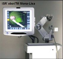
The standard method for diagnosing prostate cancer is a fine needle biopsy to remove tissue for histological examination. This is done via the rectum, guided by ultrasound and with antibiotic protection. But biopsies that result in no pathological findings, even in cases where tumors are strongly suspected, are not uncommon. The reason for this is that tumors grow in areas of the prostate that are barely accessible or not visible using rectal ultrasound. This results in repeat biopsies, because the PSA levels and DRE (digital rectal exam) results continue to be suspicious. In recent years, other imaging techniques for diagnosing prostate cancer have been established, such as magnetic resonance imaging (multi-parametric MRI) and positron emission tomography (PET/CT). The recently introduced PSMA-PET/CT has led to a significant improvement in non-invasive diagnosis of prostate cancer. This utilizes the fact that prostate cells carry prostate specific membrane antigens (PSMA) on their cell surfaces, which are easily detected and displayed using PET. These methods provide, along with transrectal ultrasound, additional evidence for the localization of a prostate tumor. If the image information from an MRI or a PET/CT is combined with the image data of the transrectal ultrasound (image fusion), an extremely accurate representation of suspected prostate tumor areas should be possible. For image-guided biopsy (fusion biopsy) we use the iSR`obotTM Mona Lisa system. With the help of this robotic system, we can employ both MRI and PSMA-PET/CT for image-guided prostate biopsy.
The prostate biopsy process
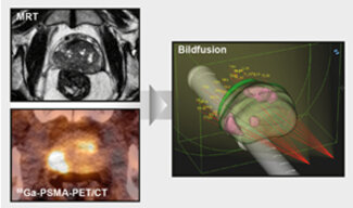
Areas where tumors are suspected from the MRI or PSMA-PET/CT scans are indicated using specialized software and then read into the biopsy device. The assessment and determination of suspected prostate tumor areas is carried out in close interdisciplinary collaboration with the Departments of Radiology and Nuclear Medicine. The digital record of an MRI or a PSMA-PET/CT, including a display of the possible tumor areas, is then fused to the real-time 3-D ultrasound image of the prostrate. Specific anatomical reference points in the prostate are used for this fusion process. After the images are merged, the doctor has a real-time 3-D ultrasound image of the prostate that includes the areas where tumors are suspected. Based on these images, in the next stage the sampling points for tissue samples can be determined. The angle and depth of penetration of the biopsy needle can be calculated by the software so that it is well positioned to access the desired areas. Through the use of "robot-assisted biopsy arms", navigation of the biopsy needle is controlled very precisely, so that optimal tissue removal from the desired site is guaranteed. By watching a screen, the operator can monitor and control the procedure at all times.
Another advantage of this system lies in the selected access path to the prostate. The increasing development of antibiotic resistance in intestinal bacteria leads over time to an increasing number of complications in transrectal prostate biopsies. In contrast, our biopsy procedure is carried out from the perineal area. An intrusion of intestinal bacteria into the bloodstream can thus be avoided.
The new tomotherapy device produces 3D images of the tumor and the adjacent region. Simultaneously, the accompanying system utilizes these images to significantly increase the precision of radiation delivery to patient tumors.
In addition, because all the tissue samples are taken via just two small entry points in the perineal area, multiple skin punctures are not necessary. Using the specialized iSR`obotTM Mona Lisa system software, localization of tissue samples can be accurately reconstructed 3-dimensionally at any time. Tissue samples of varying aggressiveness can be assigned to the sampling site, making possible a spatial representation and measurement of the prostate tumor. This information provides the basis for optimum stage-oriented treatment of prostate cancer. The surgery takes place under short anesthesia on an inpatient basis. We are happy to advise you.