Coronary Angiogram / Cardiac Catheterization

Coronary interventions
Cardiac catheterization is used to diagnose and treat diseases of the coronary arteries, heart valves and to determine heart function. Several cardiac catheter laboratories are available for examinations around the clock at the Freiburg and Bad Krozingen sites, thus ensuring the highest level of regional and supra-regional care for seriously ill patients with heart attacks or cardiogenic shock. On the other hand, we carry out planned interventions using the latest techniques and procedures and are one of the top centers in Germany and Europe in this field.
We use all modern invasive cardiology procedures and treat a wide range of patients with, for example, acute coronary syndrome (Fig. 2) or chronic total occlusions (CTO) (Fig.3). We have access to the entire spectrum of state-of-the-art materials and stent systems. The examinations are preferably performed via vascular accesses in the radial artery of the forearm. To treat complex coronary findings, we have a range of plaque-modifying procedures at our disposal, such as rotablation, intravascular lithotripsy, or orbital endarterectomy.
Implantable heart-lung machines (ECLS) or support systems (Impella) are also available at all times for high-risk interventions or for patients in cardiac arrest.
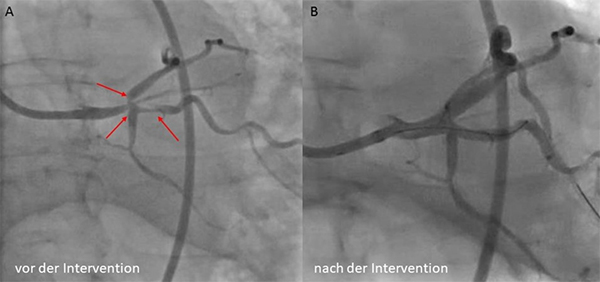
Patient with acute coronary syndrome and high-grade main stem trifurcation stenosis before (A) and after (B) treatment with DES
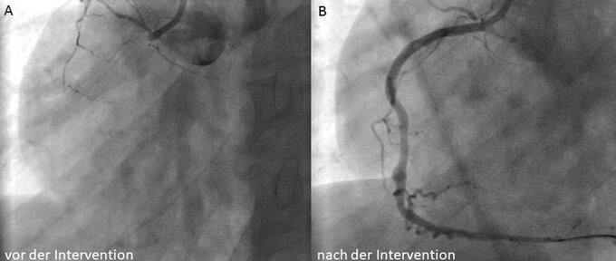
Chronically occluded right coronary artery before (A) and (B) after reopening with stenting
We also use a variety of techniques for the functional measurement and imaging of narrowing of the coronary arteries, which serve to further improve therapy and ensure optimal therapy control. These include invasive pressure measurement in the area of vascular constrictions (“fractional flow reserve measurement” = FFR; or “instantaneous flow reserve” = iFR), intravascular ultrasound (IVUS) and optical coherence tomography (OCT). This allows a detailed assessment of the vascular lumen via a small imaging catheter, which can be inserted directly via the catheter during the examination. This makes it possible, for example, to determine in a three-dimensional view whether the stent covers the entire constriction and is in good contact with the vessel wall (Fig. 4).
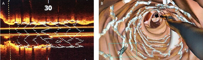
Control of optimal stent placement using OCT in the 2D view (A) and 3D reconstruction (B)