Oto-Rhino-Laryngology (ENT)

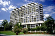
Welcome to the Department of Oto-Rhino-Laryngology of the Medical Center - University of Freiburg. Our Department provides medical assistance related to the full spectrum of Ear, Nose and Throat diseases, in particular operations on tumors in the ear, nose throat and paranasal sinus areas. Modern diagnostics and therapy, as well as ultramodern equipment, help us to create a favourable atmosphere for the treatment of all our patients.
Unique technological facilities, combined with the scientific and clinical experience and expertise of the medical staff, give us the opportunity to provide a broad spectrum of services to our patients. These include intensive tinnitus treatment, voice treatment, minimally-invasive sinus surgery, and reconstructive microsurgery of the ear to improve hearing in cases of chronic middle ear inflammations and otosclerosis.
The best results are achieved when applying innovative treatments, for example systems for navigated surgery in the head and throat area, thus increasing the precision of surgical interference to the maximum and allowing for the removal of tumors otherwise inaccessible. In addition, the application of navigational systems minimizes the risk of causing damage to adjacent tissues. The combination of systems for navigated surgery and lasers is also possible.
Exceptionally equipped in-patient wards at the Department of Oto-Rhino-Laryngology have comfortable rooms with wonderful views over the dormant volcano of the Kaiserstuhl and the French mountain range of the Vosges. A convenient and pleasant hotel atmosphere, as well as the efficiency and genuine friendliness of our staff, play an important role for the well-being of our patients.
Diagnostics and Treatment
- Microlaryngeal surgery
Using special examination instruments, the larynx and adjacent areas can be inspected through the oral cavity and anatomically preexisting upper airway, without having to make incisions from outside. In this way, wound scars and healing that would require a long hospital stay can be avoided. Under a microscope and using micromanipulators or with the help of a laser, diseased tissue can be removed or repaired without damaging the vocal cords. These procedures are performed under endotracheal or jet ventilation anesthesia. The in-patient treatment time averages 3 to 4 days.
- Diseases of throat, larynx, salivary glands, and lymph nodes
- Removal of cysts
- Tongue fraenulum and lip correction
- Anti-snoring therapy
- Parotidectomy
- Marsupialisation
- Larynx and vocal cord microsurgery
- Laser skin polishing
- Scar corrections
- Tracheoscopy
- Tonsillectomy
- Microscopic-endoscopic sinus surgery
The goal of microscopic and endoscopic sinus surgery is to create the conditions for a functional and morphological regeneration of chronically diseased mucous membranes. In this process, the existing nostrils are used for surgical access, without having to leave visible external scars. The drainage ducts which discharge from the sinuses into the nasal cavity are gently expanded and the diseased tissue removed, allowing improved ventilation and secretion drainage in order to restore the natural biological balance. These procedures are typically performed under general anesthesia. In certain cases, procedures using only local anesthesia are also possible. The in-patient hospital stay averages 4 to 7 days. Because allergies or allergy-related diseases form an important causal proportion of all chronic sinusitis cases, preoperative evaluation and treatment is carried out in close cooperation with our oto-rhino-laryngology (ear, nose & throat) allergy clinic. After surgery and hospitalization, regular follow-up care by the attending otolaryngologist is absolutely necessary while the nasal membranes regenerate.
- Paranasal sinus operations
- Removal of adenoids
- Aesthetic nose surgery
- Nose-shape correction
- Laser surgery
- Sensorineural hearing loss, acute hearing loss, acute and chronic tinnitus

Sensorineural hearing loss mostly concerns hearing loss in the inner ear, which statistically speaking becomes more common with increasing age. First, extensive diagnostics are carried out in order to determine the cause. For this we use special audiological and neurootological methods, including the latest imaging and nuclear medicine techniques available in the hospital. As far as consultation, our internist, immunologic and orthopedic colleagues are all available, along with their entire range of diagnostics, in order to initiate appropriate treatments. These may be medicinal or surgical, e.g. the implantation of hearing aids and - in close cooperation with hearing care professionals - the supply of conventional and digital hearing aids.
Acute hearing loss or acute and chronic tinnitus present an ominous clinical picture to the patient. Again, extensive diagnostics are required to clarify the possibly serious causes of these symptoms. Modern medicinal methods are generally used for treatment and, in cooperation with HBO2 - Freiburg's hyperbaric pressure chamber center - the indications for hyperbaric oxygen therapy are determined. Psychotherapeutic treatment to support the patient in changing cognitive perceptions is also initiated, in cooperation with colleagues from the established clinical fields.
- Hearing improvement microsurgery
- Inner ear surgery
Surgical inner ear interventions are indicated mainly in cases of refractory unilateral Meniere's disease. A saccotomy (intramastoidal drainage of the endolymphatic sac) is the preferred method to restore hearing. The effect of the drainage can be observed intraoperatively via electrocochleographic monitoring, in which changes in the pathologically enlarged summation potential are registered. In cases of severe unilateral inner ear hearing loss, transtympanic detachment of the saccule (Schuknecht method) can be carried out, and in certain cases a selective neurectomy of the vestibular nerve.
- All types of middle ear surgery
- Microsurgery of the middle ear
We carry out an average of 300 middle ear interventions a year, employing every microsurgical tympanoplasty technique. When indicated, reconstruction of the ossicular chain takes place using autologous, allogenic and alloplastic implants (PORP or TORP composed of various biocompatible materials, primarily titanium implants). Hearing-preserving and -enhancing middle ear surgery including stapedotomy (usually with a laser) and stapedectomy with otosclerosis are carried out under local anesthesia. Other interventions in cases of chronic mesotympanic otitis media or epitympanic otitis media and cholesteatoma are mostly performed under general anesthesia. The average hospital stay is 4 to 7 days.
- Cochlear implantation
In cases of deafness or hearing loss bordering on deafness, children and adults are supplied with electronic inner ear prostheses. Using an inner ear conserving "soft surgery" technique enables us to preserve residual hearing in about 50 percent of cases. Through a program coordinated between our Implant Center Freiburg (ICF) and related disciplines, the time needed for preliminary diagnostics is reduced to 2 to 3 days, either in-patient or out-patient. The hospital stay for surgery averages up to 7 days. Systems alignment, counselling and educational, technical and medical assistance is conducted post-operatively in the ICF. Accommodation for each 2 to 4 day stay in the ICF is in a hotel-like residence in the immediate vicinity of the treatment facility. Our implant team has more than 30 years' experience. Further details can be found on the ICF website.
- Auditory brainstem implant
The multi-channel auditory brainstem implant (ABI), which we developed, has been implanted in dozens of patients in Freiburg and Europe since 1992. It is indicated in cases of bilateral auditory nerve damage, mostly in patients with type 2 neurofibromatosis. Our close integration of otologic surgery and neurosurgery, as well as many years' experience with Cochlear implants, acoustic neuroma and cerebellopontine angle surgery, allows us to provide expert advice, diagnosis and treatment. The auditory brainstem implant is placed on the cochlear nucleus, a core area of the brain stem, to allow electrical stimulation of the auditory pathway from the lower motor neuron. Implantation is possible in patients whose primary disease is tumors or tumor recurrences, in cases of bilateral temporal bone fracture with injury to the auditory nerve, or after acoustic neuroma surgery has already taken place. With the help of this implant an acoustic connection to the environment, up to and including limited speech comprehension, is possible. Post-operative care is provided in the Implant Center Freiburg (ICF - see also Cochlear implant).
- Acoustic neuronoma
Today, thanks to audiological and imaging methods, acoustic neuronomas are usually detected early. The removal of the tumor can proceed via various access routes, depending on its size, localization and the tumor-induced hearing loss. Paramount in any surgical treatment is preservation of facial nerve function, as well as preservation of hearing in cases of suboccipital and subtemporal access, insofar as the extent of the tumor allows this. Following 1 to 5 days' preliminary assessment, all acoustic neuroma and cerebellopontine angle tumor patients transferred to the hospital are reviewed by our interdisciplinary skull base conference to determine the optimal individual treatment strategy and surgical approach. Surgery is always handled by experienced ear, nose and throat specialists and neurosurgeons. In-patient stays average 7 to 14 days.
Translabyrinthine access (through the inner ear labyrinth): About 35 to 40 percent of acoustic neuroma patients are operated on via this access route. The precondition is hearing impairment with a considerable reduction in speech intelligibility, because when using this access the inner ear will no longer function. Facial nerve function is monitored continuously during surgery.
Subtemporal access (through the middle cranial fossa on the temporal bone): This access route is used in about 15 percent of acoustic neuronomas. Aside from preserving facial nerve function, preservation of the auditory nerve and inner ear is also sought. This access is indicated for smaller auditory canal tumors where the hearing is good. Throughout the procedure, monitoring of both the facial nerve and hearing (by means of evoked potentials) is required. Intraoperative neuronavigation can be helpful in creating a targeted operation corridor.
Suboccipital access (through the rear cranial fossa): This access route is suitable for larger extrameatal cerebellopontine angle tumors and is chosen for about 55 percent of patients. Preservation of hearing is usually possible.
- Bone-anchored hearing aids
We implant bone-anchored hearing aids using the Brånemark method when indicated (bilateral microsurgically untreatable conductive hearing loss, e.g. due to malformation of the outer and middle ear or after previous remedial middle ear surgery for chronic inflammation). Sound is conducted via a retroauricular bone-anchored hearing aid, which is attached to a flunch fixing on the cranial bone via a transcutaneous spacer sleeve. The fixing and spacer sleeve are surgically implanted under local anesthesia in one or two sessions. The procedures can usually be performed on an outpatient basis.
We carry out the entire range of curative and palliative surgery of malignant and benign tumors of the ear, nose and throat area. This applies to tumors of the pharynx, oral cavity, paranasal sinuses, salivary glands, orbital cavity, larynx, base of the skull, skin of the head and neck as well as lymph vessels of the head and neck. In the field of laryngeal surgery, endoscopic and laser surgical procedures are preferred when technically feasible, but if indicated, open techniques can also be used. After extensive tumor resections, we carry out reconstruction with pedicled fasciocutaneous or myocutaneous flaps (pectoralis major flap, latissimus dorsi flap), and also by means of free microvascular anastomosed grafts (radial, vastus and upper arm flaps). In case of extensive malignancy or tumor recurrence in the oral cavity and pharynx, we employ interstitial brachytherapy (internal radiation therapy) in cooperation with the Department of Radiation Oncology.
Surgery will be performed in cases of benign and malignant tumors or trauma to the anterior, central and lateral skull base, given appropriate indications and using the most appropriate surgical access. These interventions take place after being discussed in our skull base conference, in close interdisciplinary collaboration with the specialists involved, to achieve the best possible preservation and/or enhancement of function. In planning the operation, collaboration with the neuroradiology section of the Department of Radiology is indispensable.
In addition to determining the extent and localization of the processes by means of computed tomography and/or magnetic resonance imaging, magnetic resonance angiography or digital subtraction angiography, where necessary selective embolization of the blood vessels involved - or their permanent occlusion - will be undertaken when preparing for surgery. The surgical risk and intraoperative blood loss is thus minimized, so that blood transfusions can be mostly avoided. Post-operatively, patient monitoring from an intensive care point of view is necessary. The hospital stay, depending on severity, averages 10 to 18 days.
A variety of sleep disorders can be diagnosed and treated on an out-patient basis. This depends in particular on the specific medical advice given to each individual patient. During our special sleep medicine consultation times in the university clinic, our patients can receive expert advice and help from trained sleep specialists. Given appropriate indications, a more complete diagnosis can be carried out in our ear, nose and throat sleep laboratory, where we offer sleep medicine diagnostics and therapy using the latest equipment, on an in-patient basis, by means of special cardiorespiratory polysomnography and other standardized test and treatment methods. This also includes examination of fatigue-related problems that occur in the daytime. In-patient treatment allows for the most complete medical and nursing supervision and care. The hospital stay averages 1 to 3 days. In case of special questions and findings, we enjoy close interdisciplinary cooperation with the departments of pneumology, neurology, psychiatry and pediatrics. Besides in-patient adjustment and alignment of nasal positive pressure ventilation (nCPAP), the ear, nose and throat-specific diagnosis possible in our sleep laboratory also provides analysis of surgical treatment options. These include soft palate surgery (e.g. UPPP, Uvulaflap, laser and high-frequency therapies), and nasal and sinus surgery up to so-called multilevel surgery.
Standard surgical procedure involves the use of free grafts, local or regional advanced and transposition flaps for covering defects of the skin and mucosa after resection of small benign or malignant tumors. Elaborate reconstruction measures in the head and neck area may be undertaken in case of corresponding tumor expansion. These include pedicled myocutaneous flaps (e.g. pectoralis major, deltopectoral, temporalis and latissimus flaps), free flaps, microvascular anastomosis lower and upper arm flaps for covering defects in the oral cavity and pharynx, and in some cases jejunum transplant pharynx reconstruction as part of a pharyngolaryngectomy. The jejunum transplant is performed in cooperation with the hospital's abdominal surgery department. A variety of functional-plastic, plastic-aesthetic and plastic-reconstructive surgeries of the outer ear, nose and face are frequently performed. Correction of prominent ears in preschool age children is performed on an out-patient basis using mask or endotracheal anesthesia. More difficult corrections in children and adults with auricular dysplasia usually take place on an in-patient basis. Use of the body's own rib cartilage is often necessary when rebuilding complex malformations and deformities. This is widely employed in complex rhinoplasty, for example following injuries in children with malformed external, internal or preoperated noses. In addition to the plastic reconstruction of the skin and aesthetic portions of the face and neck after tumor surgery, we also perform various surgical procedures for the rehabilitation of facial paralysis. Superficial scars can be corrected with the help of laser technology.
A CO2 laser is routinely used to perform endoscopic-microscopic surgery on benign and malignant tumors in the oral cavity, pharynx and larynx. Other deformations such as synechiae (adhesions) of the larynx are also treated with the laser. Due to its lower wavelength, the Nd:YAG laser is preferred for use on smaller vascular lesions in the nose. Larger vascular tumors are - as far as possible - embolized and then removed in the neuroradiology department. Face and nose operations are usually performed on an out-patient basis, otherwise a hospital stay of 3 to 10 days is required, depending on the disease localization and extent.
In our hospital's allergy clinic, we diagnose and treat a constantly increasing number of allergic diseases. Skin tests, challenge tests and the determination of specific immunoglobulins usually allow a clear diagnosis to be made. In-patient diagnosis and treatment may be required for complex allergic and pseudo-allergic reactions, special indications, or in patients at increased risk of severe anaphylaxis.
The treatment of inflammatory diseases in the ENT area is another large domain in our clinical spectrum. This ranges from acute tonsillitis, otitis media, furuncle of the nose, facial erysipelas and shingles (e.g. zoster oticus, ophthalmic zoster) to life-threatening infections. We have extensive experience in the treatment of deep throat abcesses, descending mediastinitis up to and including skull base osteomyelitis.
Bone-anchored epitheses are used when indicated for cosmetic rehabilitation of soft tissue defects of the nose, orbital cavity or outer ear, or after resection of malignant tumors. Anchoring of the epithesis takes place using the Brånemark method, usually in two sessions under local anesthesia. Here, titanium brackets are implanted transcutaneously on the adjacent cranial bone. Qualified anaplastologists from experienced epithesis institutions take care of adjusting the mounting device and removable epithesis in order to achieve optimal cosmetic results. Due to the expense of this procedure, a declaration is required from the relevant health insurance company in regard to accepting the costs.
Trauma includes injuries to the ear, skull base, nose and paranasal sinuses, outer throat, salivary glands as well as the larynx, trachea, pharynx and oral cavity. After appropriate diagnosis, we carry out treatment of these injuries via the necessary access routes. First, the appropriate diagnostic imaging is conducted in collaboration with the departments of diagnostic radiology and neuroradiology. Treatment also includes osteosynthetic procedures in the area of the coronal, midface and malar (cheek) bones. We treat injuries of the orbital cavity and optic nerve in close cooperation with the Departments of Ophthalmology and Oral and Maxillofacial Surgery.
Статистика
- Cochlear implantations per year: 185 patients with 202 implants (since bilateral implantations).
- Nasal septum per year: 362 patients
- Tumors (malignant) per year: 461patients
- Tumors (good o. dignity n.o.s.) per year: 957 patients
- Parotid gland: 220 patients


